Category Archives: Gene

Tongue-in-Cheek

20080216225147!Rolled_tongue_flikr

Source: Hmwith / Wikimedia Commons

Can you roll your tongue? I can. Most people can. It’s often presented as an example of a genetic trait, but don’t be fooled. It’s possible for maternal twins to differ in their tongue rolling abilities, even though maternal twins are supposed to be genetically identical [1]. Myth busted.

Sorry to disappoint. This post isn’t about the tongue rolling gene (rolled oral factor lingua, or ROFL). Maybe a few genes combine to give us the power to make the tongue tube, possibly genes that offer better muscle control or flexibility. The tongue is a weird muscle too—wiggling around in there, fiddling with that rough spot on your tooth—and it achieves that wiggling and fiddling thanks to this week’s gene of interest: myosin or MYH1.

Muscles can only pull, never push [2]. Think about that for a second. We can achieve so many artful and dexterous movements (fencing, the tango, punching someone in the face) and manage it entirely by a timely contraction of a series of tissues to achieve the desired effect. To me, that’s mind blowing. Even pushing achieved through pulling.

Push by Pulling

Muscles pull by way of two proteins, actin and myosin, that work together in a chemical tug-of-war [3]. Actin is the rope and myosin is the hand that pulls on that rope.  Myosin attaches to actin and (using energy from ATP) pulls on actin sliding it forward. Actomyosin

Muscle is organized into groups of parallel actin and myosin fibers called sarcomeres [3].  Each muscle has millions of sarcomeres—millions upon millions of molecules pulling against one another to create force.

Pitting muscles against each other, antagonistic pairs, gives us movement. For example, the bicep and the tricep (Did you get your tickets to the gun show? flexes bicep). In the tongue, several antagonistic pairs are woven together, both parallel and perpendicular to the surface. They pull against each other in a soft mass, with anchor points in the jaw, mandible, and the front and back of the throat [4]. And a few of those pairs combine to pull off the tongue roll. What a weird thing to have inside our mouths.  Enjoy being aware your tongue for the next hour!


References
[1] “Debunking the Biggest Genetic Myth of the Human Tongue“. PBS. 5 August 2015.
[2] “Why Do Muscles Only Pull And Not Push“. ezinearticles.com. 13 August 2012.
[3] “How Muscle Works“. How Stuff Works. 11 April 2001.
[4] “Anatomy Angel: The Tongue and Balance“. Dr. Dooley Noted. 30 October 2014.

Tagged , , , , , ,

Mammals Go Viral

So far on Gene of Interest, we’ve explored what can go wrong: how genes are lost; terrible diseases caused by small mutations; tiny errors that have massive consequences.  But that’s only half the picture. With all this chaos, how did humanity end up with a working genome in the first place? Where do genes come from? The likely answer: many many happy accidents, but I’d like to examine one – without it none of us would have been born – this week’s gene of interest: syncytin (ERVW-1).

Humans are mammals – we’re hairy, warm-blooded, and the ladies have boobs – and we’re part of the largest subset of mammals, placentals. All placentals have the unique ability to carry their developing young inside a womb until fully developed. A baby needs nutrients to develop, so an unborn placental has to collects nutrients from its mother’s bloodstream, but it has to do so carefully. Not only can mother and child have different blood types (so direct connection of blood vessels would be a huge no-no), to the mother’s immune system, the developing baby is a foreign invader. The mother’s white blood cells are adapted to slip themselves between other cells to reach any part of the body, so baby has to create a barrier to protect itself from mommy [1].

davincifetus

Studies of Embryos by Leonardo da Vinci

The solution is a placenta – a vast network of intertwined capillaries from both mother and child. To keep out the mother’s immune system, the cells lining the fetal side of the placenta merge into one giant cell, leaving no gaps for white blood cells to squeeze through. This single cell layer is called a syncytiotropoblast, created by the production of syncytin [2].

But syncytin is an odd gene. It’s produced nowhere else in the body, only a particular type of cell in the placenta and then never again after birth. And it’s related to a protein found in retroviruses – a protein that helps viruses invade our cells [3].

We’ve discussed retroviruses before. A retrovirus inserts its viral RNA into the host cell’s genome using an enzyme called reverse transcriptase [4].  The host cell then reads these genes like an instruction manual to build more viruses. A gene similar to syncytin allows the virus to incorporate it’s membrane with that of the cell – sort of like a smaller soap bubble merging with a larger one.

Millions of years ago, an early mammal was infected with a virus that had this syncytin gene. After the infection had subsided, the syncytin gene was left in the mammals genome and it somehow found a way to put it to work, merging one cell with another cell to create a better barrier for the placenta [5].

Syncytin

Retroviral infection with syncytin. 1) Virus invades the cell using syncytin to merge with the cell membrane. 2) Reverse transcriptase converts viral RNA into DNA. 3) The syncytin gene integrates into the cells genome. 4) The cell copies the syncytin gene. 5) The cell produces its own syncytin and uses it to merge with surrounding cells to form a syncytiotrophoblast.

According to DNA analysis, this adoption of viral DNA happened more than once. The human syncytin gene is similar to that of other primates, but completely unique from the syncytin genes found cats, mice, and rabbits. So far, scientists have found six different versions of the syncytin gene in related species of placentals, suggesting multiple ‘infection’ events and a huge selective advantage to retaining this gene [6]. So even though the placenta developed first without viral DNA, the addition of syncytin was so beneficial that everyone starting doing it.

An estimated 8 percent of our genome came from viruses – the spoils of war after an infection. Most of our viral DNA is in pieces and sits dormant, but a few are functional and even essential to our survival [7]. Odd to think that pregnancy functions thanks to an ancient viral infection. Definitely a happy accident.


References
[1] “Placenta ‘Fools Body’s Defences’“. BBC. 10 November 2007.
[2] Musicki B, Pepe G, Albrecht E. (1997). “Functional differentiation of placental syncytiotrophoblasts during baboon pregnancy: developmental expression of chorionic somatomammotropin messenger ribonucleic acid and protein levels.” J Clin Endocrinol Metab 82 (12): 4105–10, PMID 9398722
[3] “Virus Gene Syncytin Insinuated Itself in Mammalian DNA Millions of Years Ago“. SciTechDaily.com. 18 February 2012.
[4] Kurth, Reinhard; Bannert, Norbert, eds. (2010). Retroviruses: Molecular Biology, Genomics and Pathogenesis. Horizon Scientific. ISBN 978-1-904455-55-4.
[5] “The Syncytin Gene: Viruses Responsible for Human Life“. IScienceMag. 10 June 2015
[6] “Mammals Made by Viruses“. Discover Magazine. 14 February 2012.
[7] “Our Inner Viruses: Forty Million Years in the Making“. National Geographic. 1 February 2015.


Tagged , , , ,

Cyanide and the Cell

safety-44439_1280In Ian Fleming’s novels, all 00 agents are issued cyanide capsules in case of enemy capture, but James Bond threw his away. What a dummy. Sure, Bond always escapes, but why risk it? The rest of his peers understood their orders and were ready to die to keep their country’s secrets. Cyanide pills aren’t mere tropes either, but a real spy tool – a gruesome last resort to keep information out of enemy hands [1]. A lethal dose of cyanide kills in minutes – so effective because it blocks the body’s ability to make energy, via this week’s gene of interest: ATP synthase gamma subunit (ATP5C1).

There are few molecules so important that they’re found in all cellular life. DNA for one (obviously), but also the common fuel source for all cells: adenosine triphosphate or ATP [2]. Related to DNA, ATP is an adenine nucleotide with three phosphate groups attached. Think of each ATP molecule like a tiny rechargeable battery. The bonds connecting each phosphate group store the energy that drives the majority of biochemical reactions in the body – from charging neurons to powering muscle movement [3,4].

The charging stations for these molecular batteries are the mitochondria. Metabolites like glucose are broken down inside the mitochondria, one chemical bond at a time, to release energy [5]. ATP synthase, an enzyme embedded in the mitochondria, uses this released energy to make ATP. The gamma subunit (ATP5C1) sits in the center of ATP synthase and rotates to change the shape of the enzyme.  For each rotation, ATP synthase adds a phosphate group to ADP (adenosine diphosphate) to make ATP [6].  The cell then distributes these newly minted ATP molecules to power nearly all of its biochemical reactions.

ATPsyn

ATP synthase in action.  ADP and a phosphate group (pink) attach to one of three sections of ATP synthase.  As the gamma subunit (black) rotates, ATP synthase changes shape, combining ADP and the phosphate group into ATP.  ATP synthase then ejects the ATP molecule (red) and the cycle repeats [6].

ATP synthase works constantly to keep up with the cell’s energy demands. Each molecule of ATP is created and consumed at a rate of approximately 3 times per minute [7]. In all its cells, the human body contains only 250 grams of ATP, but each day it turns over an equivalent of its own body weight in ATP. At any moment, one cell can have up to one billion ATP molecules, but that’s only enough energy to keep the cell running for a few minutes [2].

Cyanide pills take advantage of the cell’s lack of energy storage. By binding to another enzyme found in mitochondria, it blocks the cell’s ability to transfer energy from glucose to ATP synthase.  No energy means no rotation of the gamma subunit inside ATP synthase, which means no new ATP. Unable to recycle spent molecules, the body burns through its ATP reserves in minutes.  It’s effectively like an off-switch for a spy – not a pretty way to go, but effective at keeping state secrets out of the hands of villains like Goldfinger.

Bond


References
[1] “Last hero of Telemark: The man who helped stop Hitler’s A-bomb“. BBC. 25 April 2013.
[2] “Nature’s batteries’ may have helped power early lifeforms“. Science Daily. 25 May 2010.
[3] “ATP and Muscle Contraction“. Boundless.com. 7 January 2016.
[4] “Nerve Impulse Transmission within a Neuron: Resting Potential “. Boundless.com. 8 January 2016.
[5] “How Cells Obtain Energy from Food“. (2002) Alberts B, Johnson A, Lewis J, et al. Molecular Biology of the Cell. 4th edition. New York: Garland Science.
[6] Nakamoto RK, Scanlon JAB, Al-Shawi MK. (2008).”The Rotary Mechanism of the ATP Synthase” Arch. Biochem. Biophys. 476 (1): 43–80.  PMID 18515057
[7] Kornberg, A. (1989) “For the love of enzymes.”Harvard University Press. Cambridge, MA.Humphery, N. ISBN-13: 978-0674307766.
[8] “How cyanide affects the electron transport chain“. The Biochem Synapse. 13 April 2013.


Tagged , , , ,

Do-It-Yourself Vitamin C

CitrusPeople can’t live without vitamin C, but everyone’s heard a story about that one college student who tried to live off of nothing but chips and ramen [1]. And were it not for a mutation a few millions of years ago, the story wouldn’t end in the clinic with a diagnosis of scurvy.  We could synthesize all the vitamin C we need with a few repairs to this week’s gene of interest: L-gulono-γ-lactone oxidase or GLO.

Vitamin C has a several important jobs. It’s an antioxidant – neutralizing nasty free-radicals [2].  It stimulates the immune system, possibly by acting as an oxidizer to destroy bacteria and viruses during infection [3].  And it’s required to make collagen, a protein found in nearly every tissue in the body [4].

Vitamin C deficiency leads to scurvy, a disease forever linked to pirates and sailors. On lengthy sea voyages, fresh fruits and vegetables didn’t last long. Without any vitamin C, sailors became sluggish, their gums would bleed, old wounds would ache, then jaundice, fever, convulsions, and finally death [5]. A horrible way to go and easily remedied by an orange or two. Multiple times in history citrus was identified and dismissed as a cure for scurvy.  Many were confused why sucking on a lime might cure scurvy, but the juice was less effective and preserved fruit even less so. Vitamin C degrades in light and heat, so processing and preservation would make the food useless. It wasn’t until the 1930’s that vitamin C was isolated and identified as the cure to scurvy.

Raw meat can even cure scurvy, because most animals (like plants) make their own vitamin C.  Humans are among a minority of animals – along with guinea pigs, bats, and other primates – that can’t synthesize their own vitamin C.  Most animals convert glucose into vitamin C using several specialized enzymes [6].  We lost the ability due to mutations in the gene for GLO, the enzyme responsible for the last step in vitamin C synthesis [7].

We still have the GLO gene, it just doesn’t work anymore.  Ever since that first mutation knocked out GLO gene function 61 million years ago, more mutations have eaten away at the gene.  Every gene is made up of several sections of coding DNA (called exons) separated by sections of non-coding DNA. Only the exons are used to build a protein. Of the 12 exons in the GLO gene, humans have lost 7 [7].

GLO gene

Human GLO pseudogene compared to the functional GLO gene from a rat [7].

GLO is an example of a pseudogene – a gene that’s lost its function.  It differs from other mutated genes we’ve discussed because it’s harmless, as long as we eat enough vitamin C to compensate. Early primates may have lost the GLO gene because their diet was rich in vitamin C. Gorging on fruits and veggies, there was no real disadvantage to loosing natural vitamin C production [6].

Our genome is likely full of psuedogenes. Each one sits dormant in our genome, a relic of our evolution. For example, scientists have identified 390 different olfactory genes, responsible for our sense of smell, but there are another 468 olfactory pseudogenes [8]. Compared to what we’ve lost, our nose is basically worthless. Think of all the smells we’re missing.

Big NoseSo that’s why you should eat your fruits and veggies – because evolution has failed you. But considering the American diet, it’s probably a good thing we can’t make our own vitamin C. If not for a horrifying death by scurvy, too many people would opt to avoid anything green for tasty, tasty junk food.


References
[1] “Scurvy Is a Serious Public Health Problem”. Slate. 20 November 2015.
[2] Padayatty SJ, Katz A, Wang Y, Eck P, Kwon O, Lee JH, Chen S, Corpe C, Dutta A, Dutta SK, Levine M. (2003). “Vitamin C as an antioxidant: evaluation of its role in disease prevention”. J Am Coll Nutr 22 (1): 18–35. PMID 12569111
[3] Wintergerst ES, Maggini S, Hornig DH.(2006) “Immune-enhancing role of vitamin C and zinc and effect on clinical conditions”. Ann Nutr Metab 50(2):85-94. PMID 16373990
[4] Peterkofsky B (1991). “Ascorbate requirement for hydroxylation and secretion of procollagen: relationship to inhibition of collagen synthesis in scurvy”. Am. J. Clin. Nutr. 54 (6): 1135S–1140S. PMID 1720597
[5] “Scott and Scurvy”. Idlewords.com. 6 March 2010.
[6] “Plagiarized Errors and Molecular Genetics”. Talkorigins.org. 5 May 2003.
[7] Drouin G, Godin JR, Pagé B. (2011). “The Genetics of Vitamin C Loss in Vertebrates”. Curr Genomics. 12 (5): 371–378. PMCID PMC3145266
[8] “The Smell of Evolution”.  National Geographic. 11 December 2013.

Tagged , , , ,

Royal Disorder

Rare genetic disorders are hard to study because they’re rare.  With public awareness almost nil, funding doesn’t come easy so treatments develop slowly.  But a few ‘lucky’ disorders present in notable patients, boosting awareness and jump-starting research.  Hemophilia is one of the lucky few, and it’s caused by a mutation in this week’s gene of interest: coagulation factor IX or F9.

Leopoldalbany

Prince Leopold, Duke of Albany

‘Royal’ hemophilia first appeared in Prince Leopold, the son of Britain’s Queen Victoria.  A delicate boy, Leopold  bruised and hemorrhaged at the slightest bump or scratch, and these wounds would take longer than normal to heal – a result of poor coagulation [1].  Little was known about hemophilia at the time, but physicians tried everything to treat the disease.  While some treatments made sense – like applying ice and compressing the wound to slow bleeding – others were a little more creative – like treating the wound with lime, hydrogen peroxide, or diluted snake venom.  If bleeding was severe, they would also transfuse hemophiliacs with the blood of a relative – a potential disaster if their blood was incompatible [2].  Ultimately, hemophiliacs like Leopold were destined to live short, sheltered lives as they could bleed to death from nearly any injury.

Queen Victoria tried to cover up Leopold’s condition, likely swearing his physicians to secrecy to protect the dignity of the bloodline [3].  While understanding of the disease was still limited, researchers had noticed it was hereditary.  If word of Leopold’s hemophilia got out, Victoria’s other children might have trouble marrying into other royal families.  She kept it a secret as best she could and hoped the disease was isolated to Leopold.  She was wrong.

Historians now believe Queen Victoria was the source of the hemophilia mutation that spread through European royalty [3].  We now know that two of her daughters were carriers and passed the mutation on to other royal families in Spain, Germany, and Russia [1].

Carriers can have the mutation, but never suffer from hemophilia.  Normally, you need two bad copies of a gene (one on each set of chromosomes) for a genetic disorder to present.  A single bad copy makes you a carrier –  you stay healthy because of your one working copy, but you might pass on the bad copy to your children.  But hemophilia is X-linked – F9 is located on the ‘X’ chromosome.  Girls have two ‘X’ chromosomes.  Boys have only one ‘X’ and one ‘Y’.  Therefore, a girl can inherit a single bad copy of F9 and have no issue, but if a boy inherits a single bad copy, he’ll develop hemophilia [4].

Leopold

Queen Victoria’s family tree: A light gray band indicates a normal copy of the F9 gene. A red band indicates a mutated copy of F9. Alice and Beatrice were both carriers of the mutated F9 gene while Leopold, with his single faulty copy of F9, developed hemophilia [1].

Technically female hemophiliacs are possible but rare, because she would have to inherit the mutation from both parents.  In Leopold’s time, it was especially rare, as hemophiliacs usually died young.  Though his condition was a secret in life, after his death, his former physicians (forgetting their word to the Queen) published several articles detailing his life and the circumstances of his death [3].  The notoriety of Leopold’s death sparked more research into this rare disease.  Soon the missing coagulation factors were identified.  Today hemophiliacs lead relatively normal lives with prophylactic treatment (regular injections of coagulation factor IX), thanks in part to high profile victims of the disease.


References
[1] “Hemophilia: ‘The Royal Disease'”. National for Case Study Teaching in Science. 20 September 2003.
[2] “History of Bleeding Disorders”. National Hemophilia Foundation.
[3] “A Royal Shame: Prince Leopold’s Hemophilia and Its Effect on Medical Research”. Dartmouth Undergraduate Journal of Science. 22 May 2009.
[4] Rogaev, E.I.; Grigorenko, A.P.; Faskhutdinova, G.; Kittler, E.L.; Moliaka, Y.K. (2009). “Genotype analysis identifies the cause of the ‘royal disease'”. Science. 326 (5954): 817. PMID 19815722

Tagged , , , ,

Error Prone

Broken Chromosomes

Damaged chromosomes (blue arrows).  Source: Square87~commonswiki / Wikimedia Commons

The world is rough, especially on our genome.  Our DNA is regularly bombarded by sunlight, radiation, smoke and soot in the air [1].  Even our own natural metabolism creates chemical by-products that can damage DNA [2].  This damage occurs anywhere from 10,000 to 1,000,000 times per cell per day – a rate that should leave us squishy, mutated blobs.

Luckily, we have repair genes regularly fixing every nick in our genome.  One such repair gene is this week’s gene of interest: xeroderma pigmentosum, complementation group A or XPA.

Antioxidants have worked their way into every fiber of the health food market.  Anti-aging, cancer-fighting superfruit supplements.  Ads claim these products protect against free-radicals – villains robbing us of our health and youth [3].  But when put to the test, few antioxidant-rich foods can live up to marketing claims.  Basically, a healthy diet has all the antioxidants we need [4].

But there’s a bit of truth to the hype.  Free-radicals are destructive.  They’re natural by-products of our metabolism that regularly damage DNA through harmful chemical reactions.  For example, one by-product, reactive oxygen species (ROS), can react to convert the DNA nucleotide guanine into 8-oxoguanine [5].
8-oxoguanine
The extra oxygen on the 8-oxoguanine isn’t a huge problem on its own, but when the cell copies its DNA, 8-oxoguanine can accidentally pair with adenine.  The code in DNA works under a very simple premise: A is paired with T, and C with G.  Therefore, if 8-oxoguanine is mispaired with adenine (G with A), that chemical reaction has effectively rewritten a small part of the genome [6].

This problem is as old as DNA – these types of chemical reactions have always eroded the code.  Nearly all life on earth has a way of fixing these errors quickly before they get out of hand.  One method is nucleotide excision repair (NER), and it requires XPA.

NER

Nucleotide Excision Repair (NER)

First, DNA is scanned for errors.  When an 8-oxoguanine base is detected, several proteins, including XPA, form a complex at the site of the error.  The protein complex unwinds the double helix and cuts the DNA several base pairs away from the 8-oxoguanine base.  Once the chunk with the error is removed, a new protein complex rewrites the missing piece of DNA [7].

Good as new!  For about 9 seconds until another error forms somewhere else.

XPA is named after xeroderma pigmentosum (XP), a disorder caused by mutations in NER genes.  Cells have other repair mechanisms besides NER, but losing a gene like XPA means no NER and errors pile up quickly, especially in the sun.  In an earlier post, I mentioned sunburns are actually skin cells killing themselves in the face of overwhelming DNA errors.  People with XP sunburn in minutes – errors build up that quickly without NER.  They are also 10,000 times more vulnerable to skin cancer [8].  It’s the loss of NER in this disorder that reveals the daily attack on our DNA.   Without it, people with XP have to live their lives in the dark, protecting their genome from the harsh rays of the sun.


References
[1] Lodish, H.; Berk, A.; Matsudaira, P.; Kaiser, CA.; Krieger, M.; Scott, M.P.; Zipursky, S.L.; Darnell, J. (2004). Molecular Biology of the Cell. New York, NY: WH Freeman. pp. 963. ISBN-10: 0-7167-3136-3.
[2] De Bont, R.; van Larebeke, N. (2004) “Endogenous DNA damage in humans: a review of quantitative data”. Mutagenesis 19 (3): 169-185. Review. PMID 15123782
[3] “POM-boozled: Do health drinks live up to their labels?”. CNN Health. 27 October 2010.
[4] “The Truth about Antioxidants”. Time. 6 August 2013.
[5] Kanvah, S.; et al. (2010). “Oxidation of DNA: Damage to Nucleobases”. Acc. Chem. Res. 43 (2): 280–287. PMID 19938827
[6] Cheng, K.C.; Cahill, D.S.; Kasai, H.; Nishimura, S.; Loeb, L.A. (1992). “8-Hydroxyguanine, an abundant form of oxidative DNA damage, causes G→T and C→A substitutions”. J Biol Chem. 267 (1): 166–72. PMID 1730583
[7] Le May, N.; Egly, J.M.; Coin, F. (2010). “True lies: the double life of the nucleotide excision repair factors in transcription and DNA repair”. J Nucleic Acids. 616342 PMID 20725631.
[8] “For Children with XP Gene, Sunlight Can Kill”. Everyday Health. 17 October 2012.


Tagged , , ,

One Man’s Junk is Another Man’s Genome

I mentioned in an earlier post that sections of our genome have no genes.  After the Human Genome Project finished in April 2003, we were left with a list of about 20,000 genes [1].  But that only accounted for 2% of the genome.  That leaves 98% that’s non-coding – 98% of our DNA that’s not a gene – seemingly without a function [2].

This result was confusing and a little disappointing.  For years, scientists assumed these sections were worthless noise – ‘junk DNA’, but the junk was passed down through generations.  Why would life bother to keep worthless DNA?  Even more confusing, complex organisms have more junk DNA.  Humans have 98%, Flies have 83%, while bacteria have only 12% [3].  If it’s useless, why do we have so much of it?

Percentage non-coding

As complexity of an organism increases, the percentage of non-coding sequences within their genome increases [3].

It turns out, these non-coding regions are not junk.  They have a few important functions, and one of those functions can be illustrated by this week’s gene of interest, paired box 6 or PAX6.

A project called ENCODE, Encyclopedia of DNA Elements, tried to decipher these non-coding sequences by cataloging every bit of the genome.  They found that, instead of 2%, closer to 80% of the genome did something.  But instead of encoding genes, non-coding DNA regulated each gene [4].  There are roughly 3 million sites in non-coding regions where proteins can stick to DNA.  Each of these sites are like switches – proteins bind to a site to turn a gene on or off.

Some of these proteins are called transcription factors – PAX6 is one of them.  It’s part of a family of Pax genes that are master regulators of important structures like the eyes [5].  PAX6 directly binds to sections of DNA right before a gene (called a promoter sequence).  Certain genes can only be expressed if PAX6 binds, just like an on/off switch.

It’s actually a little more complicated.  To turn on any gene, a minimum of 6 proteins need to attach to the promoter sequence.  Minimum of 6, but more can be added to increase or modify expression.  With up to 20 unique protein binding sites in the surrounding non-coding regions, each gene is controlled by its own complicated locking mechanism [6].

Puzzle Lock
When we first sequenced the human genome, many people thought it would read like a manual.  These non-coding sequences confused things – it seemed like the manual was filled with nonsense.  But these non-coding sequences are closer to an actual manual than the genes themselves.  The genes are the parts, but the non-coding sequences are the instructions on how to put those parts together.

Instruction Manual


References

[1] “The Human Genome Project Completion: Frequently Asked Questions””.  NIH. 30 October 2010.
[2] “Junk DNA – Not So Useless After All”.  Time. 6 September 2012.
[3] Mattick, J. S. (2007). “A new paradigm for developmental biology”. J Exp Biol 210 (9): 1526-47. PMID 17449818
[4] “Getting to Know the Genome”.  The Scientist. 5 September 2012.
[5] Fernald, R. D. (2004). “Eyes: variety, development and evolution”. Brain Behav. Evol. 64 (3): 141–7. PMID 15353906
[6] “Mysterious Non-Coding DNA – ‘Junk’ or Genetic Power Player?”.  PBS. 7 November 2011.

Tagged , , , ,

On the Wings of Mutants

insect wings 002

artwork by Molly Fischer

Molly has decided to share more of her art!  I told her I’d take anything I can get and build a post around it.  Her newest subject is insect wings, butterfly and dragonfly.  I love the detail in her work – much more flair than I have the time or the ability to do.

At first glance, this one seems tricky – what do insect wings have to do with the human genome?  Actually, a particular set of insect wings lead to the discovery of this week’s gene of interest: NOTCH1.

Back in the 1910’s, Columbia University had a room that was filled with flies [1].  A few would buzz around rotten bananas hanging from the ceiling, but most were kept in milk bottles with their newly hatched grubs.  Professor Thomas Hunt Morgan established the ‘fly room’ in 1911 to study the chromosomal theory of heredity – the theory that chromosomes are responsible for passing traits from parent to offspring.  He chose fruit flies (drosophila melanogaster) because they breed quickly (a new generation each week), they’re cheap to maintain, and they’re small, but not so small that you need a microscope to see them [2]

Morgan would expose the flies to mutagens and check each generation for mutations, noting how those traits passed down from generation to generation.  Some developed odd eye color or legs where their antennae should be.  Genes were named after these striking mutations.  For example, the gene sonic hedgehog was, in part, named after a mutant fly covered in small pointy projections resembling the quills of a hedgehog [3].

One set of flies developed oddly shaped wings.  Under the microscope, one of Morgan’s students noticed notches at the ends of each wing.  They named this mutation (and later it’s associated gene) NOTCH [4].

Notch WingNOTCH is a cell-cell receptor, which means it sits on the surface of the cell (like other cell receptors we’ve discussed), but instead of interacting with a free-floating growth factor or hormone, it connects to proteins found on the surface of another cell.  Humans have four NOTCH genes.  All four associated receptors control cell growth and are therefore important regulators of shape.  In blood vessels, NOTCH receptors regulate cell growth to control vessel diameter and branching [5].  That’s likely why a few of the veins in the notched wings are a little thicker than normal.

The ‘fly room’ uncovered many genes that are important in humans and flies alike, earning Morgan and his team a Nobel Prize in 1933 [2].  More than 0ne hundred years later, fruit flies and their mutants are still an important scientific model organism.  With thousands of mutations, and more still being generated, you’d think by now one of the mutant flies would have super powers.

Cyclops Fly


References

[1] “Columbia University Fly Room”.  Nature.com. 2014.
[2] “Thomas H. Morgan – Biographical”.  NobelPrize.org. 7 Feb 2016.
[3] Nüsslein-Volhard, C.; Wieschaus, E. (1980). “Mutations affecting segment number and polarity in Drosophila”. Nature 287 (5785): 795–801. PMID 6776413
[4] de Celis, J.F.; Garcia-Bellido, A. (1994). “Roles of the Notch gene in Drosophila wing morphogenesis”. Mech Dev 46 (2): 109-22. PMID 7918096
[5] Siekmann, A.F.; Lawson, N.D. (2007) “Notch signalling limits angiogenic cell behaviour in developing zebrafish arteries” Nature  445 (7129): 781-4. PMID 17259972

Tagged , ,

Finding Genome

Gene of Interest now has an index!  Complete with links to all eight gene posts.  That’s about 0.04% of the entire human genome.  At this rate, I’ll have discussed the entire human genome by sometime in November 2522…  So enjoy it while it lasts.

My brand-new index includes each gene’s cytogenetic location, so I thought I’d make a quick post to explain what that is.

Before the development of modern genetics, we could still study DNA by staining chromosomes and organizing them into a karyogram.  I made one of these back in high school.  You start with a print-out that looks something like this:

Karyotype_color_chromosomes_white_background

Then you cut out each chromosome, match up all 22 pairs (plus the x’s for girls or x/y for boys), and paste them on a clean sheet of paper like this:

Karyotype (cutouts)

My inner neat freak finds this so satisfying.  And this organization makes it easy to spot chromosomal abnormalities, like trisomy 21 (Down syndrome) which is caused by a third copy of chromosome 21 [1].

The bands on each chromosome are made by staining with Giemsa stain [2].  Chromosomal regions rich with adenine and thymine base pairs (AT-rich) stain darker in Giemsa stain. Regions rich with guanine and cytosine (GC-rich) stain lighter.  However, most genes are found in these lighter-stained, GC-rich regions – very few genes are found in the darker regions.  Which begs the question: if there are almost no genes in these darker regions, what’s that DNA for?  (More on that in a later post.)

These Giemsa-stained bands are useful in pinpointing the physical location of a specific gene inside a chromosome, or its cytogenetic location.  Using STAT4 as an example gene, let’s find its position on a chromosome.   Its cytogenetic location is 2q32.2.

The first number indicates which chromosome (1-22) contains the gene.  If the gene were located on one of the sex chromosomes, it would start with an ‘X’ or a ‘Y’ instead [3].

The letter ‘q’ refers the arm on which the gene can be found. Chromosomes are divided into two sections by a constriction called the centromere.  The ‘p’ refers to the shorter arm while ‘q’ refers to the longer arm [3].

The next set of numbers refers to the region and band position, respectively.  These positions are counted out from the centromere.  The number increases with increasing distance from the centromere.  Numbers after the decimal point refer to sub-band locations [3].  (In the example above, STAT4 is located on the 2nd sub-band of the 2nd band in region 3 of the long arm of chromosome 2).

Chromosome 2

Source: U.S. National Library of Medicine

These locations are identical for everyone – everybody has the same gene in the same spot.  That may seem obvious, but I think there’s something comforting about that universal organization.  To me it means, despite all our differences, we’re really a lot alike.


References

[1] Patterson, D.; (2009). “Molecular genetic analysis of Down syndrome.” Human Genetics 126 (1): 195–214. PMID 19526251.
[2] Nussbaum, R.; McInnes, R.; Willard, H. (2015). Thompson & Thompson, Genetics in Medicine (Eighth ed.). Canada: Elsevier Inc. p. 58. ISBN 978-1-4377-0696-3.
[3] “How do geneticists indicate the location of a gene”.  Genetics Home Reference. 25 January 2016

Tagged , ,

The Bones of Harry Eastlack

While playing with his sister, 5-year-old Harry Eastlack broke his leg.  He was taken to the hospital to set the fracture, but it never healed properly.  Then, at age 10, Harry’s knee and hip joints stiffened. Strange lumps formed in his thigh muscle – lumps of bone.  When surgeons removed these lumps, new bone growths appeared in their place, larger and more extensive than before.  By age 20, all of the vertebrae in Harry’s spine had fused into one piece and his most of his back muscles had ossified into boney plates.  Harry’s muscles, tendons, and ligaments were slowly turning into bone [1].  He suffered from an extremely rare disorder, called fibrodysplasia ossificans progressiva or FOP, caused by a mutation in this week’s gene of interest: activin receptor-like kinase-2 or ALK-2.

FOP is the body’s repair mechanism gone awry.  Normally injuries are repaired by regenerating wounded tissue. Tear a muscle, new muscle regrows in its place.  But people with FOP ‘heal’ connective tissue (muscle, tendons, and ligaments) by converting the wounded tissue into bone, spontaneously or upon injury [2].  Even small wounds, like a needle prick from an injection, can form tiny bone spurs under the skin.  FOP sufferers dread bumps or falls – a car crash would be a nightmare – because any injury might be ‘healed’ by bone growth that permanently locks the affected joints in place.  Even without injury, the disease progresses through the body from top to bottom, starting at the neck, then shoulders, arms, around the ribs, and finally the legs and feet [3].  Over time, each joint turns to bone, locking limbs at awkward angles and slowly converting a person into a living statue.

Again, bone formed in FOP isn’t new bone growth, but conversion of normal tissue into bone.  While that sounds pretty disturbing, it’s not a completely abnormal process, but takes its roots in normal bone development.

As I mentioned in an earlier post, bone is a living tissue, with the potential to grow, adapt, and heal itself.  There are two ways bones form [4].  One, intramembranous ossification, forms thin plates of bone like those found in the skull.  The other, endochondral ossification, starts with a template of cartilage that’s converted into bone.  This is how most of the bones in the skeleton are formed.  A temporary cartilage skeleton develops and is replaced with bone, converting one type of tissue into another.  Exactly like FOP.

Endochondral Ossification

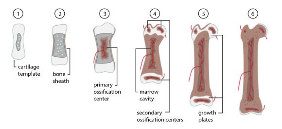
1) A cartilage template forms.  Chondrocytes (cartilage-forming cells) in the center start to die off leaving small cavities.  2) Cells around the shaft of the cartilage template convert into osteoblasts (bone-forming cells).  The osteoblasts create a sheath of bone around the template.  3) Blood vessels invade the center of the template.  Fibroblasts (wound healing cells) migrate in through the blood vessels and convert into osteoblasts.  These newly-converted osteoblasts produce bone, forming the primary ossification center.  4) The marrow cavity forms in the center.  Secondary ossification centers develop at each end.  5) The left over cartilage between ossification centers forms the growth plate, allowing the bone to grow and lengthen through adolescence.  6) The growth plate is replaced with bone in adulthood.  Cartilage remains only at the joints.

Bone growth in controlled by a set of growth factors called bone morphogenic proteins or BMPs [5].  Similar to the receptor Her2, BMP signals its bone-growth message through a set of cellular receptors that sit on the outside of the cell.  One of those receptors is ALK-2.  When BMPs interact with ALK-2, it sends pro-bone signals into the cell.

Fibrodysplasia_ossificans_progressiva.jpg

Harry Eastlack’s Skeleton on display at the Mutter Museum.  Source: Joh-co / Wikimedia Commons

In FOP, a single amino acid in ALK-2 is switched with another [6].  This mutation leaves ALK-2 in the ‘on’ position, allowing it to send pro-bone signals even without the presence of BMP growth hormones.  With this signal always on, it takes very little for cells in the body to undergo the transition similar to that found in endochondral ossification – and convert normal tissues into bone.

Much of what we know about FOP is thanks to Harry Eastlack and his skeleton, on display at The Mütter Museum of The College of Physicians in Philadelphia [1].  By the end of his life, Harry’s skeleton had fused into one solid piece.  He could only move his lips.  On his death bed, just a few days shy of his 40th birthday, he asked to donate his body to science, hoping it would help scientists understand this horrifying disorder.  The bridges and plates wrapping his skeleton like papier-mâché pointed to uncontrolled endochondral ossification.  This valuable hint helped identify the ALK-2 mutation which might eventually lead to a cure.


References
[1] “Bone, A Masterpiece of Elastic Strength”. New York Times. 27 April 2009. 
[2] van Dinther, M.; Visser, N.; de Gorter, D.J.; Doorn, J.; Goumans, M.J.; de Boer, J.; ten Dijke, P. (2011) “ALK2 R206H mutation linked to fibrodysplasia ossificans progressiva confers constitutive activity to the BMP type I receptor and sensitizes mesenchymal cells to BMP-induced osteoblast differentiation and bone formation”. J Bone Mineral Res. 25 (6): 1208-15. PMID 19929436.
[3] “FOP Symptoms”. IFOPA. 2009.
[4] Gilbert SF (2000) “Osteogenesis: The Development of Bones”. Developmental Biology. 6th edition. Sunderland (MA): Sinauer Associates.
[5] Chen, D.; Zhao, M.; Mundy, G.R. (2004) “Bone morphogenetic proteins”. Growth Factors 22(4): 233-41. PMID 15621726
[6] Shore, E.M.; Xu, M.; Feldman, G.J.; et al. (2006). “A recurrent mutation in the BMP type I receptor ACVR1 causes inherited and sporadic fibrodysplasia ossificans progressiva”. Nat. Genet. 38(5): 525–27. PMID 16642017.


Tagged , ,