Monthly Archives: February 2016

Royal Disorder

Rare genetic disorders are hard to study because they’re rare.  With public awareness almost nil, funding doesn’t come easy so treatments develop slowly.  But a few ‘lucky’ disorders present in notable patients, boosting awareness and jump-starting research.  Hemophilia is one of the lucky few, and it’s caused by a mutation in this week’s gene of interest: coagulation factor IX or F9.

Leopoldalbany

Prince Leopold, Duke of Albany

‘Royal’ hemophilia first appeared in Prince Leopold, the son of Britain’s Queen Victoria.  A delicate boy, Leopold  bruised and hemorrhaged at the slightest bump or scratch, and these wounds would take longer than normal to heal – a result of poor coagulation [1].  Little was known about hemophilia at the time, but physicians tried everything to treat the disease.  While some treatments made sense – like applying ice and compressing the wound to slow bleeding – others were a little more creative – like treating the wound with lime, hydrogen peroxide, or diluted snake venom.  If bleeding was severe, they would also transfuse hemophiliacs with the blood of a relative – a potential disaster if their blood was incompatible [2].  Ultimately, hemophiliacs like Leopold were destined to live short, sheltered lives as they could bleed to death from nearly any injury.

Queen Victoria tried to cover up Leopold’s condition, likely swearing his physicians to secrecy to protect the dignity of the bloodline [3].  While understanding of the disease was still limited, researchers had noticed it was hereditary.  If word of Leopold’s hemophilia got out, Victoria’s other children might have trouble marrying into other royal families.  She kept it a secret as best she could and hoped the disease was isolated to Leopold.  She was wrong.

Historians now believe Queen Victoria was the source of the hemophilia mutation that spread through European royalty [3].  We now know that two of her daughters were carriers and passed the mutation on to other royal families in Spain, Germany, and Russia [1].

Carriers can have the mutation, but never suffer from hemophilia.  Normally, you need two bad copies of a gene (one on each set of chromosomes) for a genetic disorder to present.  A single bad copy makes you a carrier –  you stay healthy because of your one working copy, but you might pass on the bad copy to your children.  But hemophilia is X-linked – F9 is located on the ‘X’ chromosome.  Girls have two ‘X’ chromosomes.  Boys have only one ‘X’ and one ‘Y’.  Therefore, a girl can inherit a single bad copy of F9 and have no issue, but if a boy inherits a single bad copy, he’ll develop hemophilia [4].

Leopold

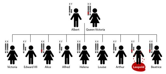
Queen Victoria’s family tree: A light gray band indicates a normal copy of the F9 gene. A red band indicates a mutated copy of F9. Alice and Beatrice were both carriers of the mutated F9 gene while Leopold, with his single faulty copy of F9, developed hemophilia [1].

Technically female hemophiliacs are possible but rare, because she would have to inherit the mutation from both parents.  In Leopold’s time, it was especially rare, as hemophiliacs usually died young.  Though his condition was a secret in life, after his death, his former physicians (forgetting their word to the Queen) published several articles detailing his life and the circumstances of his death [3].  The notoriety of Leopold’s death sparked more research into this rare disease.  Soon the missing coagulation factors were identified.  Today hemophiliacs lead relatively normal lives with prophylactic treatment (regular injections of coagulation factor IX), thanks in part to high profile victims of the disease.


References
[1] “Hemophilia: ‘The Royal Disease'”. National for Case Study Teaching in Science. 20 September 2003.
[2] “History of Bleeding Disorders”. National Hemophilia Foundation.
[3] “A Royal Shame: Prince Leopold’s Hemophilia and Its Effect on Medical Research”. Dartmouth Undergraduate Journal of Science. 22 May 2009.
[4] Rogaev, E.I.; Grigorenko, A.P.; Faskhutdinova, G.; Kittler, E.L.; Moliaka, Y.K. (2009). “Genotype analysis identifies the cause of the ‘royal disease'”. Science. 326 (5954): 817. PMID 19815722

Tagged , , , ,

Error Prone

Broken Chromosomes

Damaged chromosomes (blue arrows).  Source: Square87~commonswiki / Wikimedia Commons

The world is rough, especially on our genome.  Our DNA is regularly bombarded by sunlight, radiation, smoke and soot in the air [1].  Even our own natural metabolism creates chemical by-products that can damage DNA [2].  This damage occurs anywhere from 10,000 to 1,000,000 times per cell per day – a rate that should leave us squishy, mutated blobs.

Luckily, we have repair genes regularly fixing every nick in our genome.  One such repair gene is this week’s gene of interest: xeroderma pigmentosum, complementation group A or XPA.

Antioxidants have worked their way into every fiber of the health food market.  Anti-aging, cancer-fighting superfruit supplements.  Ads claim these products protect against free-radicals – villains robbing us of our health and youth [3].  But when put to the test, few antioxidant-rich foods can live up to marketing claims.  Basically, a healthy diet has all the antioxidants we need [4].

But there’s a bit of truth to the hype.  Free-radicals are destructive.  They’re natural by-products of our metabolism that regularly damage DNA through harmful chemical reactions.  For example, one by-product, reactive oxygen species (ROS), can react to convert the DNA nucleotide guanine into 8-oxoguanine [5].
8-oxoguanine
The extra oxygen on the 8-oxoguanine isn’t a huge problem on its own, but when the cell copies its DNA, 8-oxoguanine can accidentally pair with adenine.  The code in DNA works under a very simple premise: A is paired with T, and C with G.  Therefore, if 8-oxoguanine is mispaired with adenine (G with A), that chemical reaction has effectively rewritten a small part of the genome [6].

This problem is as old as DNA – these types of chemical reactions have always eroded the code.  Nearly all life on earth has a way of fixing these errors quickly before they get out of hand.  One method is nucleotide excision repair (NER), and it requires XPA.

NER

Nucleotide Excision Repair (NER)

First, DNA is scanned for errors.  When an 8-oxoguanine base is detected, several proteins, including XPA, form a complex at the site of the error.  The protein complex unwinds the double helix and cuts the DNA several base pairs away from the 8-oxoguanine base.  Once the chunk with the error is removed, a new protein complex rewrites the missing piece of DNA [7].

Good as new!  For about 9 seconds until another error forms somewhere else.

XPA is named after xeroderma pigmentosum (XP), a disorder caused by mutations in NER genes.  Cells have other repair mechanisms besides NER, but losing a gene like XPA means no NER and errors pile up quickly, especially in the sun.  In an earlier post, I mentioned sunburns are actually skin cells killing themselves in the face of overwhelming DNA errors.  People with XP sunburn in minutes – errors build up that quickly without NER.  They are also 10,000 times more vulnerable to skin cancer [8].  It’s the loss of NER in this disorder that reveals the daily attack on our DNA.   Without it, people with XP have to live their lives in the dark, protecting their genome from the harsh rays of the sun.


References
[1] Lodish, H.; Berk, A.; Matsudaira, P.; Kaiser, CA.; Krieger, M.; Scott, M.P.; Zipursky, S.L.; Darnell, J. (2004). Molecular Biology of the Cell. New York, NY: WH Freeman. pp. 963. ISBN-10: 0-7167-3136-3.
[2] De Bont, R.; van Larebeke, N. (2004) “Endogenous DNA damage in humans: a review of quantitative data”. Mutagenesis 19 (3): 169-185. Review. PMID 15123782
[3] “POM-boozled: Do health drinks live up to their labels?”. CNN Health. 27 October 2010.
[4] “The Truth about Antioxidants”. Time. 6 August 2013.
[5] Kanvah, S.; et al. (2010). “Oxidation of DNA: Damage to Nucleobases”. Acc. Chem. Res. 43 (2): 280–287. PMID 19938827
[6] Cheng, K.C.; Cahill, D.S.; Kasai, H.; Nishimura, S.; Loeb, L.A. (1992). “8-Hydroxyguanine, an abundant form of oxidative DNA damage, causes G→T and C→A substitutions”. J Biol Chem. 267 (1): 166–72. PMID 1730583
[7] Le May, N.; Egly, J.M.; Coin, F. (2010). “True lies: the double life of the nucleotide excision repair factors in transcription and DNA repair”. J Nucleic Acids. 616342 PMID 20725631.
[8] “For Children with XP Gene, Sunlight Can Kill”. Everyday Health. 17 October 2012.


Tagged , , ,

Weaponized Genetics

Genome editing a potential WMD?  According to the recent worldwide threat assessment, genetic engineering is as concerning as North Korea’s nuclear program.  Sounds silly, but the implications of recent advances, particularly CRISPR-Cas9, are pretty scary.  Follow the link below to my newest article on LadyFreethinker.org to read more.

Genome Editing Now Listed as a Weapon of Mass Destruction

CRISPR-Cas9 acts like a word processor for the human genome.  The potential advances in medicine, agriculture, even human evolution are staggering.  I’ve attached a great video by Youreka Science and iBiology that explains how CRISPR-Cas9 works using only a white board.  I mean the video uses a white board, not CRISPR… Check it out below.

Tagged , , ,

One Man’s Junk is Another Man’s Genome

I mentioned in an earlier post that sections of our genome have no genes.  After the Human Genome Project finished in April 2003, we were left with a list of about 20,000 genes [1].  But that only accounted for 2% of the genome.  That leaves 98% that’s non-coding – 98% of our DNA that’s not a gene – seemingly without a function [2].

This result was confusing and a little disappointing.  For years, scientists assumed these sections were worthless noise – ‘junk DNA’, but the junk was passed down through generations.  Why would life bother to keep worthless DNA?  Even more confusing, complex organisms have more junk DNA.  Humans have 98%, Flies have 83%, while bacteria have only 12% [3].  If it’s useless, why do we have so much of it?

Percentage non-coding

As complexity of an organism increases, the percentage of non-coding sequences within their genome increases [3].

It turns out, these non-coding regions are not junk.  They have a few important functions, and one of those functions can be illustrated by this week’s gene of interest, paired box 6 or PAX6.

A project called ENCODE, Encyclopedia of DNA Elements, tried to decipher these non-coding sequences by cataloging every bit of the genome.  They found that, instead of 2%, closer to 80% of the genome did something.  But instead of encoding genes, non-coding DNA regulated each gene [4].  There are roughly 3 million sites in non-coding regions where proteins can stick to DNA.  Each of these sites are like switches – proteins bind to a site to turn a gene on or off.

Some of these proteins are called transcription factors – PAX6 is one of them.  It’s part of a family of Pax genes that are master regulators of important structures like the eyes [5].  PAX6 directly binds to sections of DNA right before a gene (called a promoter sequence).  Certain genes can only be expressed if PAX6 binds, just like an on/off switch.

It’s actually a little more complicated.  To turn on any gene, a minimum of 6 proteins need to attach to the promoter sequence.  Minimum of 6, but more can be added to increase or modify expression.  With up to 20 unique protein binding sites in the surrounding non-coding regions, each gene is controlled by its own complicated locking mechanism [6].

Puzzle Lock
When we first sequenced the human genome, many people thought it would read like a manual.  These non-coding sequences confused things – it seemed like the manual was filled with nonsense.  But these non-coding sequences are closer to an actual manual than the genes themselves.  The genes are the parts, but the non-coding sequences are the instructions on how to put those parts together.

Instruction Manual


References

[1] “The Human Genome Project Completion: Frequently Asked Questions””.  NIH. 30 October 2010.
[2] “Junk DNA – Not So Useless After All”.  Time. 6 September 2012.
[3] Mattick, J. S. (2007). “A new paradigm for developmental biology”. J Exp Biol 210 (9): 1526-47. PMID 17449818
[4] “Getting to Know the Genome”.  The Scientist. 5 September 2012.
[5] Fernald, R. D. (2004). “Eyes: variety, development and evolution”. Brain Behav. Evol. 64 (3): 141–7. PMID 15353906
[6] “Mysterious Non-Coding DNA – ‘Junk’ or Genetic Power Player?”.  PBS. 7 November 2011.

Tagged , , , ,

On the Wings of Mutants

insect wings 002

artwork by Molly Fischer

Molly has decided to share more of her art!  I told her I’d take anything I can get and build a post around it.  Her newest subject is insect wings, butterfly and dragonfly.  I love the detail in her work – much more flair than I have the time or the ability to do.

At first glance, this one seems tricky – what do insect wings have to do with the human genome?  Actually, a particular set of insect wings lead to the discovery of this week’s gene of interest: NOTCH1.

Back in the 1910’s, Columbia University had a room that was filled with flies [1].  A few would buzz around rotten bananas hanging from the ceiling, but most were kept in milk bottles with their newly hatched grubs.  Professor Thomas Hunt Morgan established the ‘fly room’ in 1911 to study the chromosomal theory of heredity – the theory that chromosomes are responsible for passing traits from parent to offspring.  He chose fruit flies (drosophila melanogaster) because they breed quickly (a new generation each week), they’re cheap to maintain, and they’re small, but not so small that you need a microscope to see them [2]

Morgan would expose the flies to mutagens and check each generation for mutations, noting how those traits passed down from generation to generation.  Some developed odd eye color or legs where their antennae should be.  Genes were named after these striking mutations.  For example, the gene sonic hedgehog was, in part, named after a mutant fly covered in small pointy projections resembling the quills of a hedgehog [3].

One set of flies developed oddly shaped wings.  Under the microscope, one of Morgan’s students noticed notches at the ends of each wing.  They named this mutation (and later it’s associated gene) NOTCH [4].

Notch WingNOTCH is a cell-cell receptor, which means it sits on the surface of the cell (like other cell receptors we’ve discussed), but instead of interacting with a free-floating growth factor or hormone, it connects to proteins found on the surface of another cell.  Humans have four NOTCH genes.  All four associated receptors control cell growth and are therefore important regulators of shape.  In blood vessels, NOTCH receptors regulate cell growth to control vessel diameter and branching [5].  That’s likely why a few of the veins in the notched wings are a little thicker than normal.

The ‘fly room’ uncovered many genes that are important in humans and flies alike, earning Morgan and his team a Nobel Prize in 1933 [2].  More than 0ne hundred years later, fruit flies and their mutants are still an important scientific model organism.  With thousands of mutations, and more still being generated, you’d think by now one of the mutant flies would have super powers.

Cyclops Fly


References

[1] “Columbia University Fly Room”.  Nature.com. 2014.
[2] “Thomas H. Morgan – Biographical”.  NobelPrize.org. 7 Feb 2016.
[3] Nüsslein-Volhard, C.; Wieschaus, E. (1980). “Mutations affecting segment number and polarity in Drosophila”. Nature 287 (5785): 795–801. PMID 6776413
[4] de Celis, J.F.; Garcia-Bellido, A. (1994). “Roles of the Notch gene in Drosophila wing morphogenesis”. Mech Dev 46 (2): 109-22. PMID 7918096
[5] Siekmann, A.F.; Lawson, N.D. (2007) “Notch signalling limits angiogenic cell behaviour in developing zebrafish arteries” Nature  445 (7129): 781-4. PMID 17259972

Tagged , ,

Finding Genome

Gene of Interest now has an index!  Complete with links to all eight gene posts.  That’s about 0.04% of the entire human genome.  At this rate, I’ll have discussed the entire human genome by sometime in November 2522…  So enjoy it while it lasts.

My brand-new index includes each gene’s cytogenetic location, so I thought I’d make a quick post to explain what that is.

Before the development of modern genetics, we could still study DNA by staining chromosomes and organizing them into a karyogram.  I made one of these back in high school.  You start with a print-out that looks something like this:

Karyotype_color_chromosomes_white_background

Then you cut out each chromosome, match up all 22 pairs (plus the x’s for girls or x/y for boys), and paste them on a clean sheet of paper like this:

Karyotype (cutouts)

My inner neat freak finds this so satisfying.  And this organization makes it easy to spot chromosomal abnormalities, like trisomy 21 (Down syndrome) which is caused by a third copy of chromosome 21 [1].

The bands on each chromosome are made by staining with Giemsa stain [2].  Chromosomal regions rich with adenine and thymine base pairs (AT-rich) stain darker in Giemsa stain. Regions rich with guanine and cytosine (GC-rich) stain lighter.  However, most genes are found in these lighter-stained, GC-rich regions – very few genes are found in the darker regions.  Which begs the question: if there are almost no genes in these darker regions, what’s that DNA for?  (More on that in a later post.)

These Giemsa-stained bands are useful in pinpointing the physical location of a specific gene inside a chromosome, or its cytogenetic location.  Using STAT4 as an example gene, let’s find its position on a chromosome.   Its cytogenetic location is 2q32.2.

The first number indicates which chromosome (1-22) contains the gene.  If the gene were located on one of the sex chromosomes, it would start with an ‘X’ or a ‘Y’ instead [3].

The letter ‘q’ refers the arm on which the gene can be found. Chromosomes are divided into two sections by a constriction called the centromere.  The ‘p’ refers to the shorter arm while ‘q’ refers to the longer arm [3].

The next set of numbers refers to the region and band position, respectively.  These positions are counted out from the centromere.  The number increases with increasing distance from the centromere.  Numbers after the decimal point refer to sub-band locations [3].  (In the example above, STAT4 is located on the 2nd sub-band of the 2nd band in region 3 of the long arm of chromosome 2).

Chromosome 2

Source: U.S. National Library of Medicine

These locations are identical for everyone – everybody has the same gene in the same spot.  That may seem obvious, but I think there’s something comforting about that universal organization.  To me it means, despite all our differences, we’re really a lot alike.


References

[1] Patterson, D.; (2009). “Molecular genetic analysis of Down syndrome.” Human Genetics 126 (1): 195–214. PMID 19526251.
[2] Nussbaum, R.; McInnes, R.; Willard, H. (2015). Thompson & Thompson, Genetics in Medicine (Eighth ed.). Canada: Elsevier Inc. p. 58. ISBN 978-1-4377-0696-3.
[3] “How do geneticists indicate the location of a gene”.  Genetics Home Reference. 25 January 2016

Tagged , ,